There's a method of looking at the brain, called "resting-state connectivity," a form of fMRI. fMRI indirectly estimates where areas of higher and lower brain activity are occurring. (Areas that are more active consume more glucose, and require more blood flow in order to supply them with that glucose. fMRI measures these changes in blood flow). "Connectivity" measures the co-occurrence of brain activity--which regions are active at the same time, and therefore might be working together. "Resting state" means that connectivity is being measured while a person sits still in the MRI machine, not doing any particular task. This is different from fMRI studies that look at "functional connectivity," which is the co-occurrence of activity while performing a specific task.
Resting-state activity can be hard to interpret and is still somewhat controversial. That's because people aren't doing anything in particular, they're just mind-wandering, or worrying about problems, or thinking how bored they are and wondering when the experiment will be over, or finding patterns in the scanner noise--or any number of other things. The experimenter has no control over what you're thinking or perceiving during a resting state experiment. What's amazing is that researchers can still find consistent brain networks during experiments like this. This might be because we habitually use our brains in certain ways while thinking, and these relationships exist even when we're not performing any particular task. It may also occur for mind-wandering in particular because mind-wandering is a task with its own brain network(s), just like other (better-defined) tasks.
Not surprisingly, a research group decided to compare resting-state connectivity in autistic and neurotypical adults. You can find an abstract here and a summary for non-scientists here. (The research team was Marlene Behrmann's group at Carnegie Mellon, which does a lot of autism research and is especially interested in perception). The group was trying to address conflicting results about whether autistic brains are "underconnected" or "overconnected." Some studies find less connectivity in autistic than neurotypical brains, others find greater connectivity. It's not clear whether the conflicting results come from different study methods, artifacts (i.e., autistic people move more in the scanner), heterogeneity in the autistic population, or some other factor. My personal theory was that in addition to heterogeneity, the explanation comes from connectivity in autism being atypically high in some brain areas or networks, and atypically low in others, and that furthermore the pattern varies based on what task a person is doing. So, whether you find overconnected or underconnected autistic brains depends on what you're having people do, and what parts of the brain you're looking at.
Behrmann's group specifically recruited autistic adults with IQ in the normal range, perhaps because this made it easier to explain the task and ensure participants stayed still in the scanner. They looked at connectivity both between the brain's left and right hemisphere, and within each of these hemispheres.
As I would have predicted, they found that some areas were more highly connected in the autistic group and other areas were less connected.
But here's where it gets interesting:
"Connectivity patterns were pretty similar between neurotypical participants. Their brains all looked roughly the same. By contrast, there was a LOT of variability in the ASD group. Connectivity patterns were 'idiosyncratic.'"
The boring part of this finding is that participants who were rated as having stronger "behavioral symptoms of ASD" also differed more from the normal "template." Well, of course, autism is by definition different than the norm, so we shouldn't be surprised when autistic brains differ from "average" brains, or even that more autistic people should have more divergent brains. The interesting result is that autistic brains were different from each other.
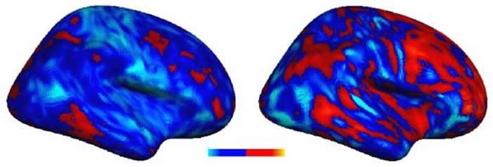
Above: Image from the described paper, accessed via the Neuroscience News writeup, which describes it as follows: "This image shows a comparison in the extent of the vowel deviation from the typical profile of two individuals with autism [a voxel is a 3d pixel that is the unit of measurement in an fMRI study]. The individual with the more severe autism symptoms (right) showed greater deviations, both positive (more red) and negative (lighter blue) from the typical inter-hemispheric connectivity pattern compared to the individual with the less severe autism symptoms (left)."
While in some cases the pattern is qualitatively similar in the two brains (e.g., increased connectivity in frontal areas and in ventral temporal areas), other regions show opposite patterns. For example, the area where the occipital lobe (back of the brain) meets the parietal lobe (top/back of the brain) has atypically low connectivity in the "less severe" brain on the left and atypically high connectivity in the "more severe" brain on the right. The same is true for the medial temporal region (just below the dark indentation in the middle of the brain).
This result seems like the neural flip side of a point Mel Baggs (Ballastexistenz/Withasmoothroundstone) often makes about behavior: for any trait you can think of, autistic people are often found at both extremes while neurotypical people are usually in the middle. You can find autistic people with extremely high and extremely low pain sensitivity, language ability, spatial ability, empathy, tendency to introspect, and so on. Not only are their abilities extreme and often opposite of each other, but so, it turns out, are their patterns of brain connectivity. We need to remember "heterogeneity" can mean not just "variable" but sometimes "opposite," both for behavior and brains.
No comments:
Post a Comment